Cataract Surgery: What Happens During the Procedure
Dr. Guillermo Enríquez TostadoNovember 15, 20269 min read

In this article
Cataract surgery involves removing the cloudy natural lens and replacing it with an intraocular lens. Although it is usually performed with local anesthesia and through small incisions, the exact steps may vary depending on the eye’s anatomy, the type of cataract, other eye conditions, and the lens selected. This article explains, in general terms, what may happen before and during the procedure, without replacing an assessment by an ophthalmologist.
What happens during cataract surgery?
A cataract is clouding of the natural lens, which helps focus light on the retina. When vision declines and begins to affect everyday activities, an ophthalmologist may consider surgery. The procedure aims to remove the cloudy natural lens and place an intraocular lens, also called an IOL. The National Eye Institute notes that surgery is the only way to remove a cataract.
Not everyone needs surgery at the same time. The decision depends on how much the cataract affects vision and daily life, as well as on individual risks and benefits. If cataracts are present in both eyes, each eye is usually treated on a separate occasion, according to the specialist’s plan.
Preparation and anesthesia
Before surgery, measurements of the eye are taken to calculate the power of the intraocular lens. These tests help select the implant best suited to the eye’s length and shape, although they cannot guarantee that the final vision will be exactly as expected. It is also important to share information about medications, general health conditions, previous eye surgeries, and other vision problems.
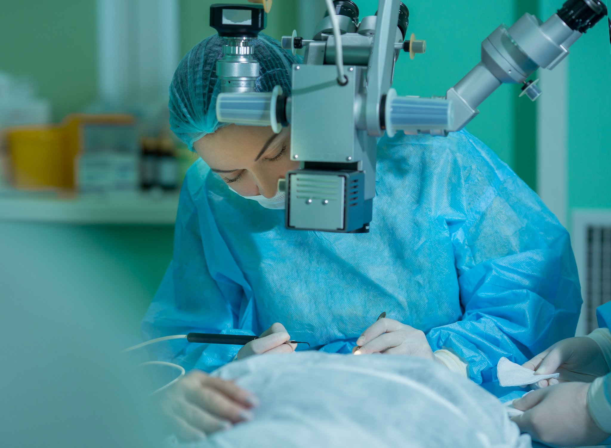
In many cases, surgery is performed with local anesthesia. This may involve anesthetic eye drops, an injection around the eye, or a combination of techniques, depending on the clinical situation and the surgical team’s practice. The American Academy of Ophthalmology, through EyeWiki, describes topical anesthesia as one of the most commonly used forms for cataract surgery in the United States; in some cases, it may be supplemented with anesthetic inside the eye.
With local anesthesia, the person is usually awake. They may perceive lights, movement, or changes in brightness, but normally cannot see in detail what the surgeon is doing. In some situations, medication is used to help reduce anxiety. General anesthesia is not usually used and may be considered in specific circumstances, for example, when a person cannot remain still or tolerate a local technique; this decision is made by the medical team.
General steps of the procedure
The steps may vary depending on the technique, the condition of the eye, and the surgeon’s experience, but generally include:
- 1.Preparing the eye: the pupil is dilated, the skin around the eye is cleaned, and a sterile drape is placed, leaving the eye to be operated on exposed.
- 2.Accessing the inside of the eye: the surgeon makes one or more small incisions in or near the cornea. Their size and location depend on the technique and the needs of the case.
- 3.Opening the capsule: the natural lens is accessed through its capsule, a transparent structure that surrounds it. In many procedures, the surgeon aims to preserve the back portion of this capsule to support the intraocular lens.
- 4.Fragmenting and removing the lens: the most common technique uses an ultrasound probe to break up the cloudy natural lens and remove it by suction. This procedure is known as phacoemulsification. The description from the National Health Service explains that ultrasound may be used to break up the natural lens before removing it.
- 5.Implanting the lens: once the natural lens has been removed, the intraocular lens is placed inside the eye, usually in the capsule that held the natural lens.
- 6.Review and discharge: the team checks the eye before the person goes to recovery. Many surgeries are performed as outpatient procedures, although the total time at the facility may be longer than the time spent in surgery itself.
Traditional surgery is usually performed with small instruments and ultrasound. Laser-assisted surgery is also available and uses a laser for some steps. The American Academy of Ophthalmology explains that these are different techniques and that the choice should be discussed with the surgeon based on the patient’s individual characteristics. Using a laser does not mean that everyone will have the same outcome or that the risks of surgery.
The intraocular lens: an individual decision
The intraocular lens remains inside the eye to replace the optical function of the removed natural lens. Selection takes into account eye measurements, astigmatism, the health of the cornea and retina, the person’s activities, and their visual expectations.
- •Monofocal: What it aims to offer: A primary focus at one distance, often distance vision. Points to discuss: You may need glasses for reading or other distances.
- •Toric: What it aims to offer: Help correct corneal astigmatism in selected patients. Points to discuss: Its effect depends on the measurements, placement, and stability of the lens.
- •Multifocal or extended-range: What it aims to offer: Distribute focus across more than one distance and reduce, for some patients, dependence on glasses. Points to discuss: Visual effects such as halos or glare may occur; it is not suitable for everyone.
Intraocular lenses have different focusing objectives. The professional labeling for the SBL-3 multifocal lens, available from the FDA, describes a specific device; it is not a general guide for patients and does not establish that it is used in this practice. A lens with more than one focusing distance does not guarantee that you will no longer need glasses. A monofocal lens may be suitable depending on visual priorities and eye health.
What can vary from one case to another?
Surgery is not performed exactly the same way in every eye. The cataract’s hardness or location, pupil size, eye shape, astigmatism, previous eye surgery, glaucoma, diabetes, or retinal diseases may affect preparation, anesthesia, technique, or lens type.
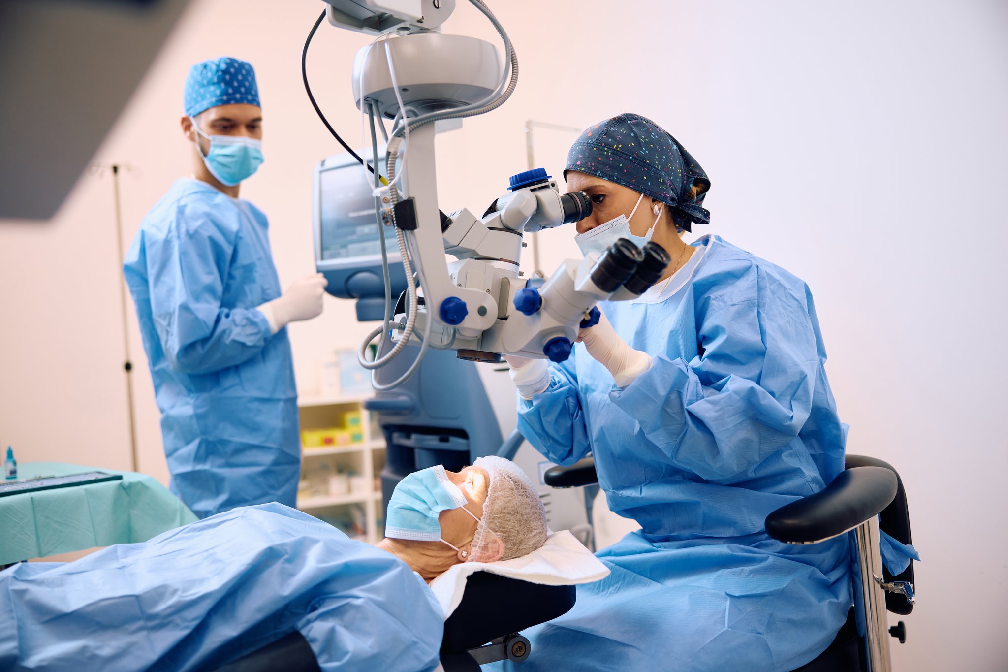
In some cases, the surgeon may need to use additional instruments, enlarge an incision, place stitches, or modify the initial plan. These situations do not necessarily mean the procedure was performed incorrectly; they reflect that surgery may require decisions during the procedure. That is why an explanation beforehand should include alternatives, limitations, and risks, not just the ideal steps.
After surgery, temporary blurred vision, light sensitivity, itching, or mild discomfort may occur. The care team will explain how to protect the eye and which drops to use. The National Eye Institute advises seeking medical attention right away for vision loss, persistent severe pain, marked redness, flashes, or a significant increase in floaters.
Like any procedure, surgery has risks, including infection, inflammation, bleeding, changes in eye pressure, retinal detachment, double vision, or vision loss. An opacity of the posterior capsule, known as a “secondary cataract,” may also develop months or years later and can be treated with laser when indicated. Individual assessment and follow-up appointments are essential.
Frequently asked questions
Will I be asleep during cataract surgery?
Usually not. Many surgeries are performed with local anesthesia, and the person remains awake, although the eye is numbed. You may notice lights or movement. The type of anesthesia depends on the case and should be explained before the procedure.
Will an intraocular lens eliminate the need for glasses?
Not necessarily. Some lenses are designed for one primary distance, while others may help you see at several distances, but no implant guarantees complete freedom from glasses. Visual needs, eye health, and possible optical effects should be discussed before choosing.
Is the removal always done with a laser?
No. The most common technique uses an ultrasound probe to break up and remove the natural lens. Laser-assisted surgery is also available, but whether it is appropriate depends on the case, and it is not universally better.
What to bring to your consultation
Understanding what cataract surgery involves can help you have a clearer conversation about anesthesia, removal of the natural lens, and choosing an intraocular lens. However, the steps, risks, and visual outcome may vary. An ophthalmology assessment can review the overall health of your eye, explain the alternatives, and set realistic expectations.
If you would like to discuss questions about your case, you can request an assessment on the cataract surgery page. The decision should be made using individual clinical information and without assuming that one technique or type of lens is suitable for everyone.
References
- •
- •
- •
- •
- •
This content is for informational purposes only and does not replace a medical consultation or constitute a diagnosis.
This information is for guidance only and does not replace a medical consultation, diagnosis, or treatment. Each case is assessed in person.


